Lower Body Skeleton Diagram - The Lower Limbs Human Anatomy And Physiology Lab Bsb 141. Consists of the skull, ribs, vertebral column and sternum. Diagram of a human female skeleton, back view. Human skeleton is a framework of our body. A diagram of the lower extremities. Female anatomy includes the external genitals, or the vulva, and the internal reproductive organs.
Drawn sleleton lower body pencil and in color drawn sleleton lower body. Thigh anatomy bones and muscles. The skeleton acts as a scaffold by providing support and protection for the soft tissues that make up the rest of the body. The appendicular skeleton totals 126 bones, consisting of the pectoral girdles, the upper limbs, the pelvic girdle and the lower limbs. Find the perfect human skeleton diagram stock illustrations from getty images.
File Human Arm Bones Diagram Svg Wikipedia from upload.wikimedia.org The lumbar region of the spine, more commonly known as the lower back, is situated between the thoracic, or chest, region of the spine, and the sacrum. The horizontal central part on each side is the body of the mandible. There are 206 bones in the body which make up the skeleton. A wooden ball on a table; This framework consists of many individual bones and cartilages. It is the strongest and most prominent part of the lower extremity, thus a personal favourite for fitness enthusiasts to showcase. The bones of the pelvis and lower back work together to support the body's weight, anchor the abdominal and hip muscles, and protect the delicate vital organs of the vertebral and abdominopelvic cavities. Let's take a look at the bones of the appendicular skeleton.
The vertebral column of the lower back includes the five lumbar vertebrae, the sacrum, and the coccyx.
The first diagram summarizes the different muscular compartments (fascial compartments) of the thigh and leg, and the different fascias (crural fascia, intermuscular septum, interosseous membrane, adductor canal, fascia lata) Folkens (2005) puts the bodyweight contributed by the bones at about 20%. It is the strongest and most prominent part of the lower extremity, thus a personal favourite for fitness enthusiasts to showcase. Now that we've learned about the hip and pelvis, we'll explore the thigh anatomy. Female anatomy includes the external genitals, or the vulva, and the internal reproductive organs. A diagram of the lower extremities. This article looks at female body parts and their functions, and it provides an interactive diagram. Stronger muscles can help stabilize the lower back and can help reduce injury risk. If the pulleys are assumed frictionless and of small. The healthy skeletal system is made up of bones, ligaments, and cartilage. The appendicular skeleton includes the bones of the shoulder girdle, the upper limbs, the pelvic girdle, and the lower limbs. The muscles of the lower back help stabilize, rotate, flex, and extend the spinal column, which is a bony tower of 24 vertebrae that gives the body structure and houses the spinal cord. The vertebral column of the lower back includes the five lumbar vertebrae, the sacrum, and the coccyx.
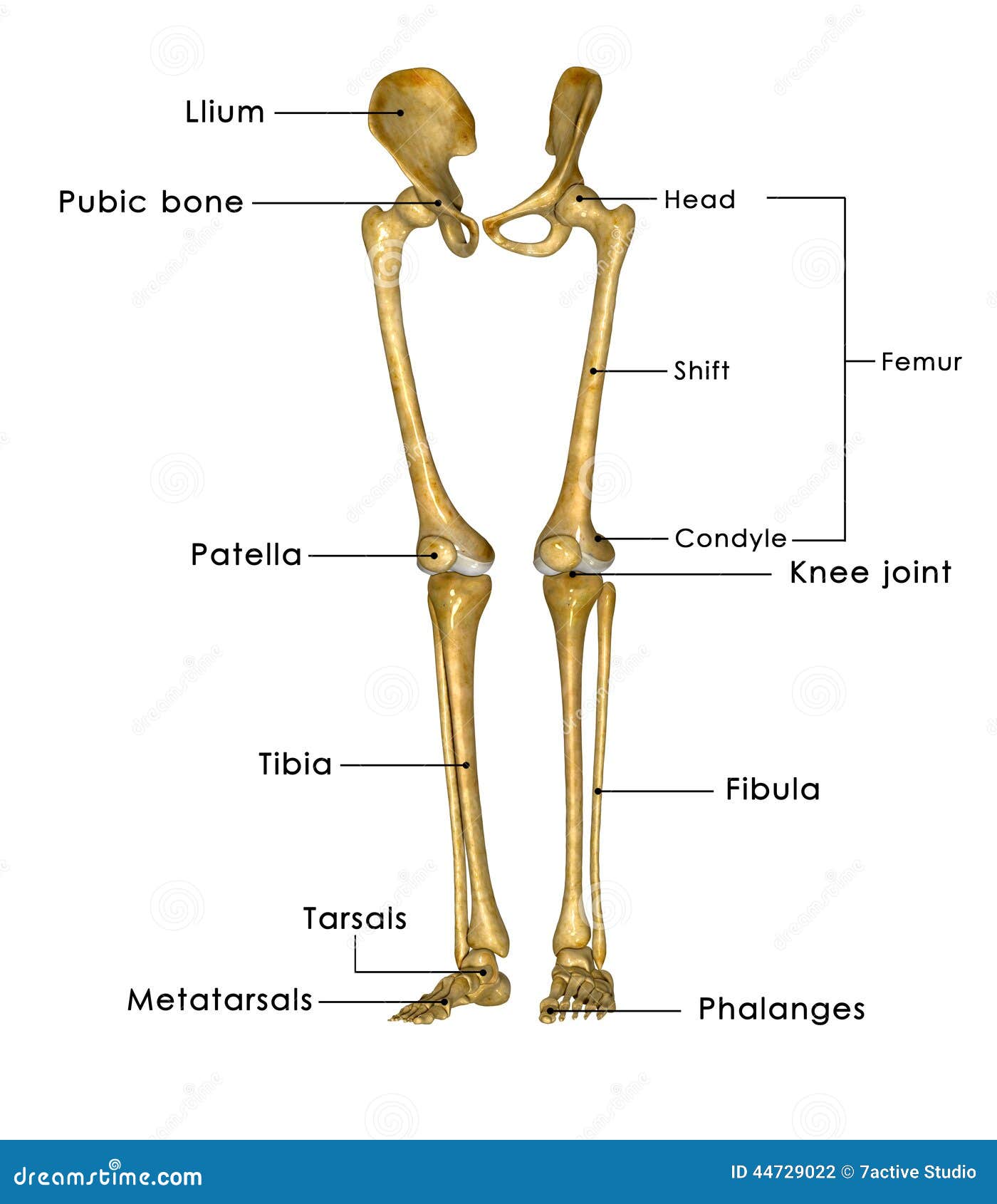
Find the perfect human skeleton diagram stock illustrations from getty images. The red lines point individual bones and the names are writen in singular, the blue lines conect to group of bones and are in plural form. It is the strongest and most prominent part of the lower extremity, thus a personal favourite for fitness enthusiasts to showcase. This article looks at female body parts and their functions, and it provides an interactive diagram. This framework consists of many individual bones and cartilages.
Skeleton Legs Stock Illustration Illustration Of Muscle 44729022 from thumbs.dreamstime.com The part of the skeleton associated with the vertical axis of the body. This article looks at female body parts and their functions, and it provides an interactive diagram. Folkens (2005) puts the bodyweight contributed by the bones at about 20%. Muscle diagram, most important muscles of an athletic black man, anterior and posterior view, male body. Drawn sleleton lower body pencil and in color drawn sleleton lower body. Hip and leg bone diagram / human body anatomy 3d illustration of hip legs and hands skeletal and cardiovascular systems viewed from the back on white background stock photo alamy. There are 5 different types of bones in the skeleton. The appendicular skeleton totals 126 bones, consisting of the pectoral girdles, the upper limbs, the pelvic girdle and the lower limbs.
Human skeleton, the internal skeleton that serves as a framework for the body.
If the pulleys are assumed frictionless and of small. Folkens (2005) puts the bodyweight contributed by the bones at about 20%. The upper portion of the body is the alveolar margin, corresponding to the alveolar margins of the. Proper posture and body mechanics. The muscles of the lower back help stabilize, rotate, flex, and extend the spinal column, which is a bony tower of 24 vertebrae that gives the body structure and houses the spinal cord. Lower body skeleton diagram / anatomy of lower extremity. Stronger muscles can help stabilize the lower back and can help reduce injury risk. Understanding the anatomy of your lower spine can help you communicate more effectively with the medical professionals who treat your lower back pain. Find the perfect human skeleton diagram stock illustrations from getty images. Muscle anatomy head 12 photos of the muscle anatomy head dog head muscle. Lower body skeleton diagram / anatomy of lower extremity. Free body diagrams are therefore drawn for the lengths ax1 and. The horizontal central part on each side is the body of the mandible.
The skeletal system includes all of the bones and joints in the body. It is the strongest and most prominent part of the lower extremity, thus a personal favourite for fitness enthusiasts to showcase. The first diagram summarizes the different muscular compartments (fascial compartments) of the thigh and leg, and the different fascias (crural fascia, intermuscular septum, interosseous membrane, adductor canal, fascia lata) Woman holding a blackboard with an illustration of the human digestive system drawn on it in chalk. Hip and leg bone diagram / human body anatomy 3d illustration of hip legs and hands skeletal and cardiovascular systems viewed from the back on white background stock photo alamy.
Skeletal System Lower Body Diagram Quizlet from o.quizlet.com The vertebral column of the lower back includes the five lumbar vertebrae, the sacrum, and the coccyx. This article looks at female body parts and their functions, and it provides an interactive diagram. There is a little difference between the male and female skeleton, but for diagrams mostly a male skeletal. Keep your spine erect and lift objects with your legs. Find the perfect human skeleton diagram stock illustrations from getty images. There also are bands of fibrous connective tissue—the ligaments and the tendons—in intimate relationship with the parts of the skeleton. Thigh anatomy bones and muscles. The horizontal central part on each side is the body of the mandible.
The skeleton is divided into 2 main parts:
The appendicular skeleton includes the bones of the shoulder girdle, the upper limbs, the pelvic girdle, and the lower limbs. Start studying skeleton lower body. The skeletal system includes all of the bones and joints in the body. Consists of the skull, ribs, vertebral column and sternum. Abdominal muscles picture anatomy 12 photos of the abdominal muscles picture anatomy abdominal muscles anatomy diagram, abdominal muscles picture anatomy, human anatomy, abdominal muscles anatomy diagram, abdominal muscles picture anatomy Thigh anatomy bones and muscles. Muscle anatomy head 12 photos of the muscle anatomy head dog head muscle. It's also the largest joint in the body. A diagram of the lower extremities. Teachme anatomy part of the teachme series the medical information on this site is provided as an information resource only, and is not to be used or relied on for any diagnostic or treatment purposes. The healthy skeletal system is made up of bones, ligaments, and cartilage. Femur the longest and the strongest bone in the human skeleton system as you can observe in the following labeled skeleton of the human body. The ilium is the big bone of the hip, the ischium is the bone on which one sits and the pubis forms the lower frontal hip bone as seen in the labeled human skeleton diagram.
Folkens (2005) puts the bodyweight contributed by the bones at about 20% lower body diagram. Animals with a skeleton (vertebrates) are a tiny minority as.
Share :
Post a Comment
for "Lower Body Skeleton Diagram - The Lower Limbs Human Anatomy And Physiology Lab Bsb 141"


Post a Comment for "Lower Body Skeleton Diagram - The Lower Limbs Human Anatomy And Physiology Lab Bsb 141"